odontogenic keratocyst removal
- Consistent with odontogenic keratocyst benign ribbon-like squamous epithelium with keratinization separated from the underlying hyaline stroma with cartilage. The most common affection region is the posterior region of the lower jaw.
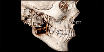
Jaw Reconstruction Surgery Orthognathic Surgery India
A provisional diagnosis of odontogenic keratocyst was made based on these findings.

. Preoperative orthopantomogram before extraction showing multilocular radiolucency in mandible with root resorption. See also Odontogenic tumours and cysts. Cysts are filled with keratinous debris.
Long-term follow-up with monitoring by X-ray is important as if these cysts are left untreated they can become quite large and locally destructive. The unique histopathological appearance and the propensity for recurrence has made it management controversial in terms of the conservatism to be followed. The posterior mandible is the most frequent site involved.
Head and neck pathology. - Benign respiratory mucosa with mild inflammation. A Common and Serious Clinical Misdiagnosis.
- NEGATIVE for malignancy. Implant surgery for the placement of dental implants is performed after full bony consolidation of the bone grafts to complete full oral rehabilitation for the patient. Download Citation Removal of Odontogenic Keratocyst in Maxilla Through the Le Fort I Osteotomy The odontogenic keratocyst is a lesion with specific clinical and histopathological aspects.
Osteotomy in the trigonoretromolar region until the exposure of the lesion. The excision of the overlying mucosa. Odontogenic keratocyst arises from the remnants of dental lamina either in the mandible or in the maxilla.
The most appropriate surgical approaches for the successful. They were surgically treated through an intraoral approach by resection without continuity defects. This 26-year-old man came to our hospital with a complaint of severe pain and swelling in his lower jaw left side.
Surgical enucleation of the lesion was planned under general anaesthesia after endodontic treatment of the involved teeth. Odontogenic keratocyst of the maxilla. Cyst can be removed by open as well as endoscopic approach.
They may also suggest getting a biopsy where you will get part of the cyst removed and sent to a laboratory for further examination. Immediate mandibular reconstruction with a corticocancellous iliac crest bone graft. Odontogenic keratocysts OKCs are benign intraosseous odontogenic lesions that have a locally aggressive behavior and exhibit a high recurrence rate after the treatment.
Odontogenic Cyst Treatment. Surgical Removal of Odontogenic Keratocyst. Enucleation of the cyst and removal of the associated teeth.
Nancy Herbst walks through how to properly remove an Odontogenic KeratocystLike and subscribe for moreUnion City Oral Surgery Group is located in Union. The principle of treatment of odontogenic. The most commonly arise from the epithelial cell rests of the dental lamina.
Treatment of the odontogenic keratocyst involves meticulous resection to completely remove the lesion followed by reconstruction of the jaw with bone grafting. J Am Dent Assoc. We recommend the following protocol in the management of large mandibular OKC.
Enucleation of the lesion. A 19-year-old male patient undergoing dental treatment presented a well-delimited hypodense image around the crown of tooth 48 included during tomographic examination. Although enucleationcurettage has the advantage over marsupialization of providing a complete specimen for histopathologic.
Ali M Baughman RA. Br J Oral Maxillofac Surg. Of a total of 227 odontogenic cysts 31 odontogenic keratocysts were histopathologically diagnosed preoperatively.
The lower border of the mandible andor the posterior border of the ramus was left intact. Removal of the cyst with removal of surrounding bone and or cryosurgery intense cold is applied to the cyst and bone are the most common forms of treatment. The presentation can be unilocular or multilocular in appearance.
However complete removal of the OKC can be difficult because of the thin friable epithelial lining. A Clinicopathologic Study of 312 Cases. To enucleate is to remove whole or clean as a tumour from its envelope Curettage is defined as the removal of growths or other material from the wall of a cavity 14Enucleation with and without various adjuncts has been utilized for many years.
Large odontogenic keratocysts sometimes are treated initially by cystotomy and insertion of a drainage tube which can promote shrinkage of the lesion and fibrous thickening of the cyst wall before subsequent total removal. CT scans in axial and coronal planes. The odontogenic keratocyst is a distinct entity arising from odontogenic epithelium.
A mild pain was present since the past few. Case Report Figure 4. Biopsy of the lesion.
Right Maxillary Sinus Mass Excision. We opted for an endoscopic approach because it offers minimal reductive change. Odontogenic keratocysts can initially be treated with incisional biopsy and decompression by installing a polyethylene drain to allow subsequent reduction of the cystic cavity size resulting in thickening of the capsule which allows a later easy removal withapparently lower relapse rate waldron.
Dental professionals will typically recommend a test like an MRI CT or X-ray. Incision on the right mandibular ramus up to the distal aspect of tooth from the buccal aspect. Odontogenic keratocysts are benign developmental cysts which cause extensive destruction of bone.
Odontogenic keratocyst are surgically removed by marsupialisation enucleation and chemical curettage with carnoys solution. In rare instances particularly large cysts may require resection and bone grafting.

Management Of Benign Odontogenic Lesions In The Paediatric Patient

Recurrence Of Odontogenic Keratocyst Okc In Relation To Cortical Download Scientific Diagram

Pdf Removal Of Odontogenic Keratocyst In Maxilla Through The Le Fort I Osteotomy Semantic Scholar

Pdf Removal Of Odontogenic Keratocyst In Maxilla Through The Le Fort I Osteotomy Semantic Scholar

Jaw Reconstruction Surgery With Bone Graft Odontogenic Keratocyst

Endoscopically Assisted Enucleation And Curettage Of Large Mandibular Odontogenic Keratocyst Oral Surgery Oral Medicine Oral Pathology Oral Radiology And Endodontics
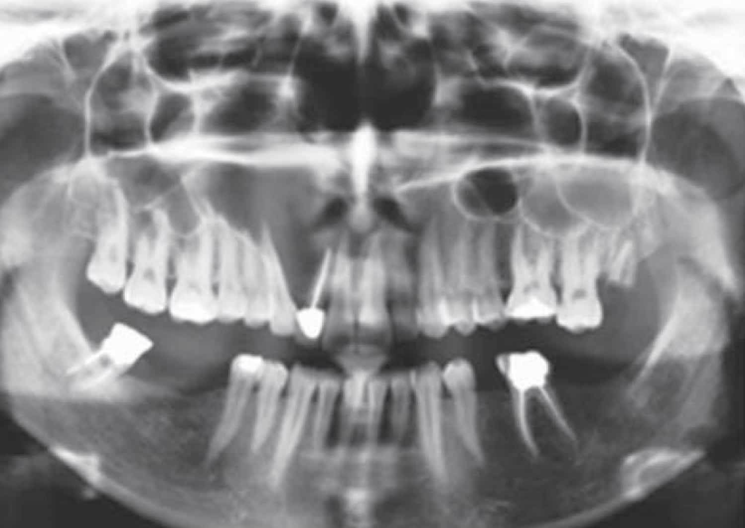
Odontogenic Keratocyst Definition Causes Symptoms Diagnosis Treatment Prognosis

Keratocystic Odontogenic Tumor Treatment Modalities Study Of 3 Cases
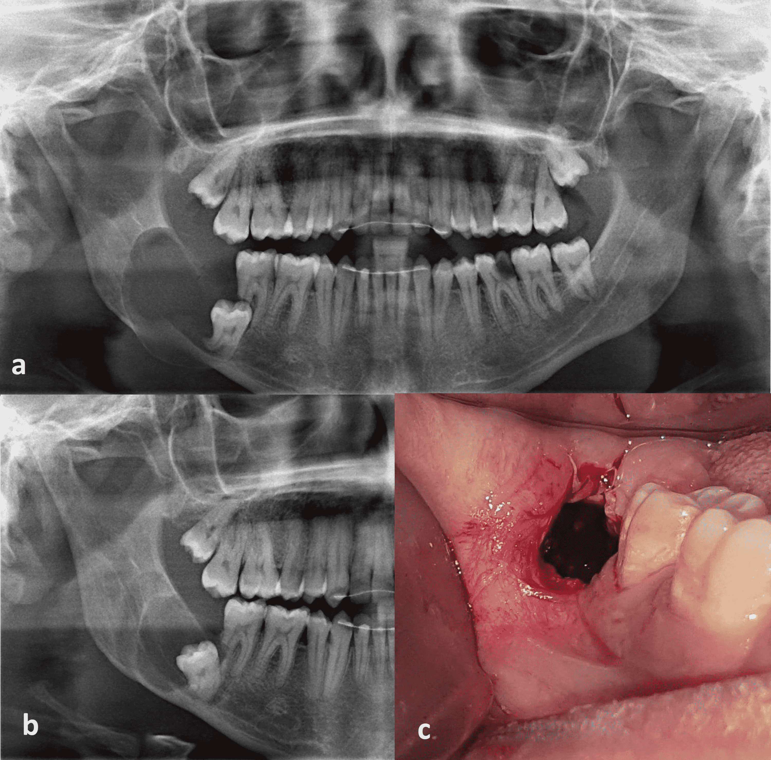
Cureus Marsupialization Of Dentigerous Cysts Followed By Enucleation And Extraction Of Deeply Impacted Third Molars A Report Of Two Cases
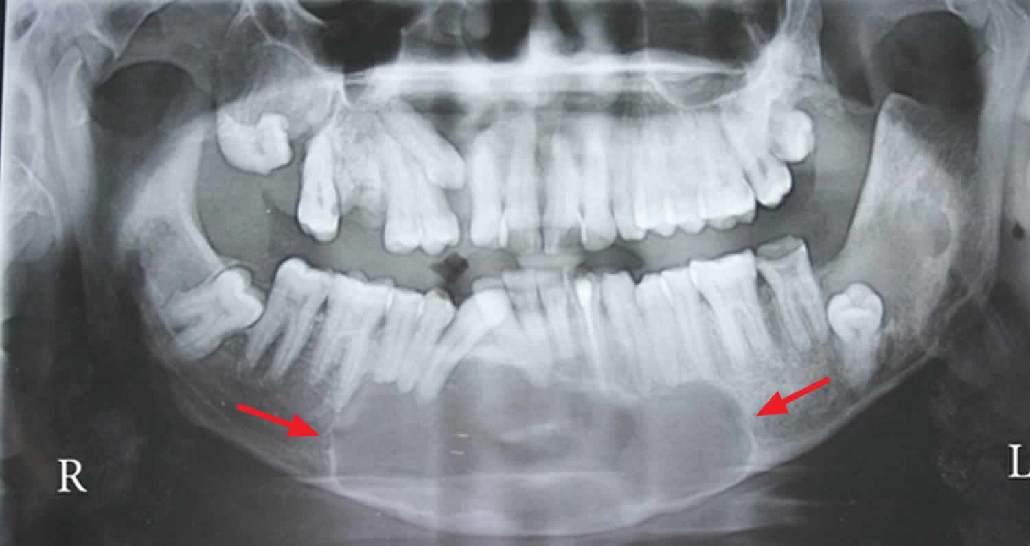
Odontogenic Keratocyst Definition Causes Symptoms Diagnosis Treatment Prognosis
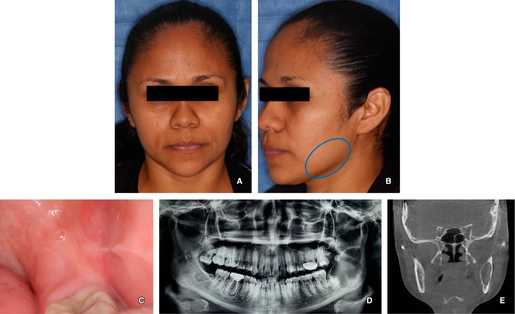
Treatment With Decompression Of An Odontogenic Keratocyst

A Orthopantomogram Shows Unilocular Odontogenic Keratocyst In Third Download Scientific Diagram

Conservative Management Of Odontogenic Keratocyst With Long Term 5 Year Follow Up Case Report And Literature Review Sciencedirect
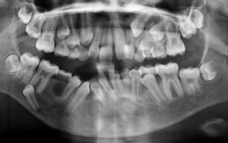
Odontogenic Keratocyst Okc Exodontia
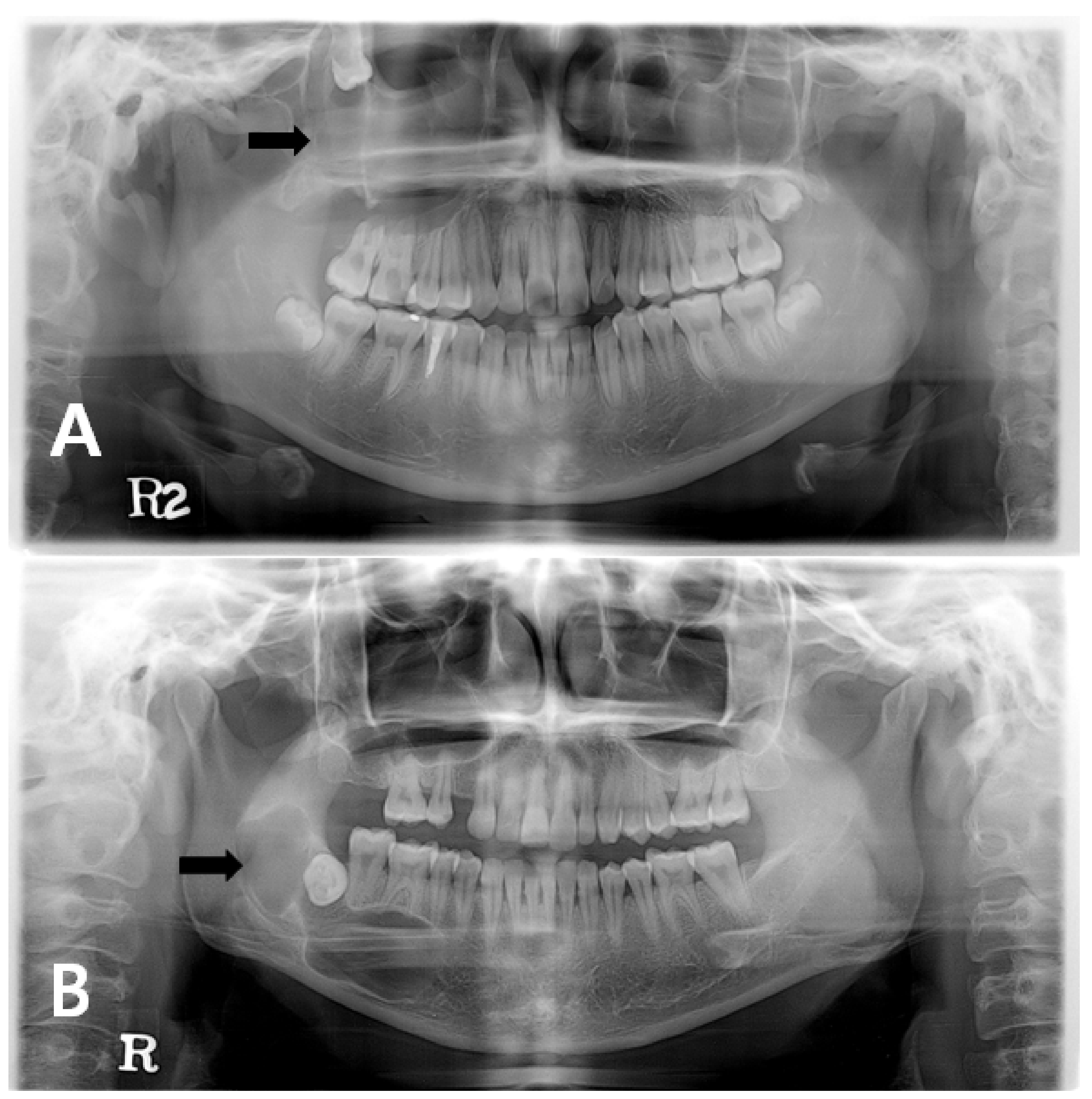
Jcm Free Full Text Changes In Cellular Regulatory Factors Before And After Decompression Of Odontogenic Keratocysts Html
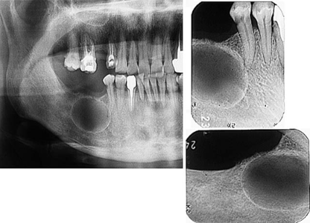
Odontogenic Keratocyst Okc Exodontia

Case Presentation Of A Patient Affected By Okc That Didn T Experience Download Scientific Diagram

Odontogenic Keratocyst Clinical Features Pathogenesis Radiographic Features Youtube
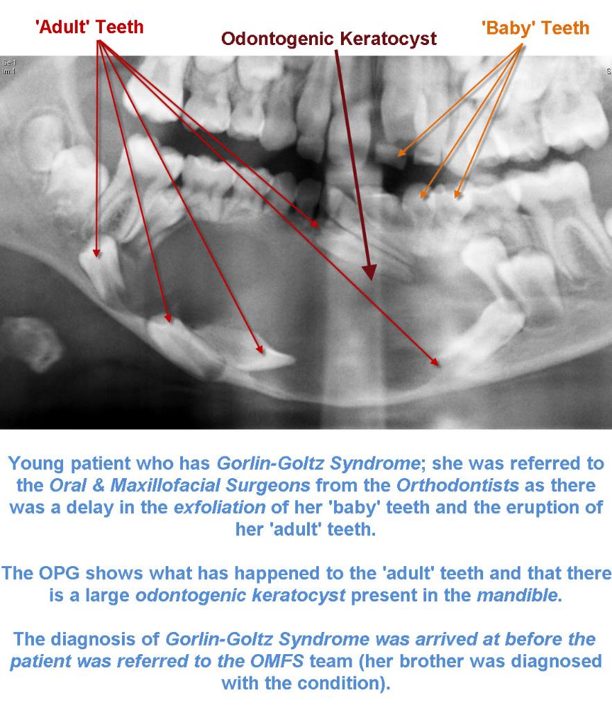
Odontogenic Keratocyst Okc Exodontia


0 Response to "odontogenic keratocyst removal"
Post a Comment